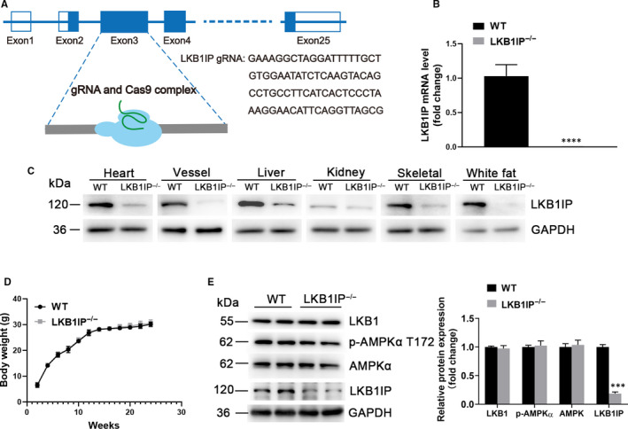
FIGURE 2.

Establishment and analysis of LKB1IP knockout mice model. (A) Schematic diagram of CRISPR/Cas9 to knock out LKB1IP. (B) Quantitative PCR analysis of LKB1IP mRNA level in the hearts of wild‐type (WT) (n = 3) and LKB1IP‐/‐ mice (n = 5). ****P < .0001 vs WT. (C) Western blot analysis of LKB1IP protein expression in tissues of WT and LKB1IP‐/‐ mice. (D) Bodyweight of WT and LKB1IP‐/‐ mice (n = 5). (E) Western blot analysis of LKB1, p‐AMPKα T172 and AMPKα protein expression in hearts of WT and LKB1IP‐/‐ mice(n = 3). ***P < .001 vs WT
