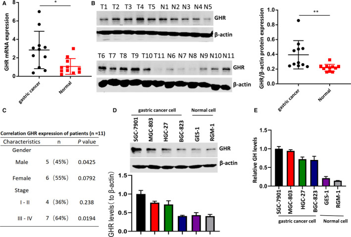
FIGURE 1.

The expression levels of GHR in gastric cancer tissues and cell lines. A, GHR was highly expressed in tumour tissues compared with normal mucosa tissues. B, The expression of GHR was examined by Western blotting in tumour tissues compared with normal mucosa tissues. C, The clinicopathologic characteristics of patients and the correlation of GHR were shown. P value was determined by Student's t test. D, GHR level was significantly increased in gastric cancer cell lines (SGC‐7901, MGC‐803, HGC‐27 and BGC‐823) compared with human gastric epithelial cell lines (GES‐1 and RGM‐1). *P < 0.05 as determined by Student's t test. E, The expression of GH in four gastric cancer cell lines and two human gastric epithelial cell line was determined by ELISA
