
Fig. 4.
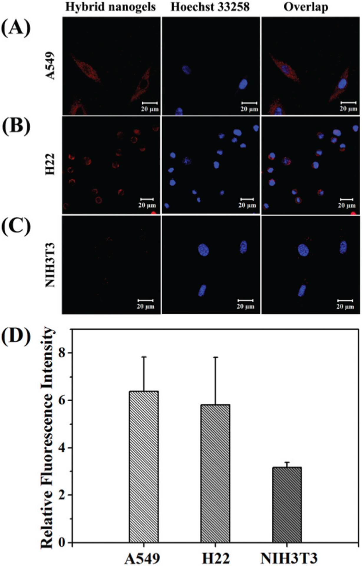
Confocal images of (A) A549, (B) H22 and (C) NIH3T3 cells after incubation with mHA-GC hybrid nanogels (red). The nuclei were dyed with Hoechst 33258 (blue). (D) Flow cytometry analysis of mHA-GC hybrid nanogels incubated with A549, H22 and NIH3T3 cells compared with the control respectively (4 h of incubation at 37 °C); data as mean values ± SD (n = 3).