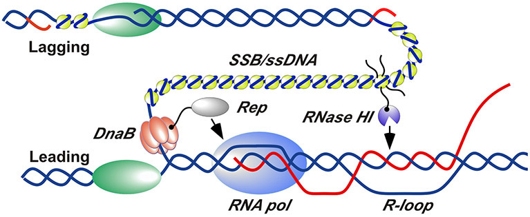
Figure 5. Schematic model for RNase HI and Rep helicase localization and action at sites of replication/transcription collision.
RNase HI (purple) is localized to the DNA replication fork by interaction with SSB (yellow). Rep helicase (grey) is localized by interaction with DnaB (orange). SSB-Ct tails are shown explicitly for only one SSB tetramer for clarity. DNA strands are shown in blue, RNA strands are shown in red, and DNA polymerases are shown in green. Several replisome components have been omitted or separated for clarity.

