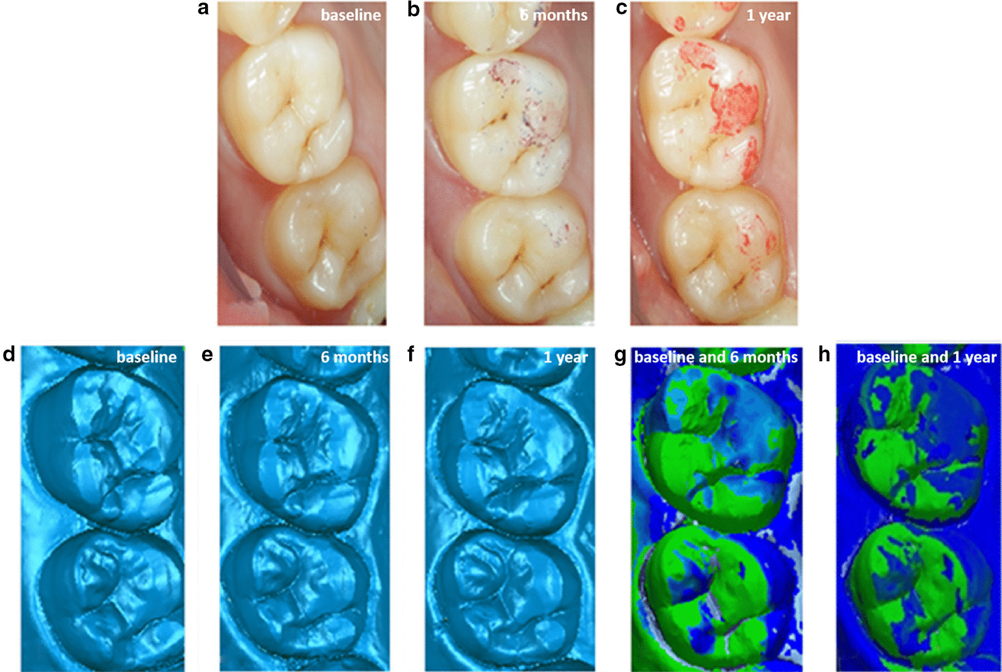
Fig. 3.

Image of different time intraoral photograph and three-dimensional morphology and overlap deviation of #16 (antagonist): a immediately after cementing of the crowns; b at the 6-month recall; c at the 1-year recall; d three-dimensional morphology immediately after cementing of the crowns; e three-dimensional morphology at the 6-month recall; f dark blue indicates three-dimensional morphology at the 1-year recall; g three-dimensional morphology immediately and at 6-month superimposed image showing wear areas; h dark blue indicates three-dimensional morphology immediately and at the 1-year superimposed image showing wear areas
