Fig. 5.
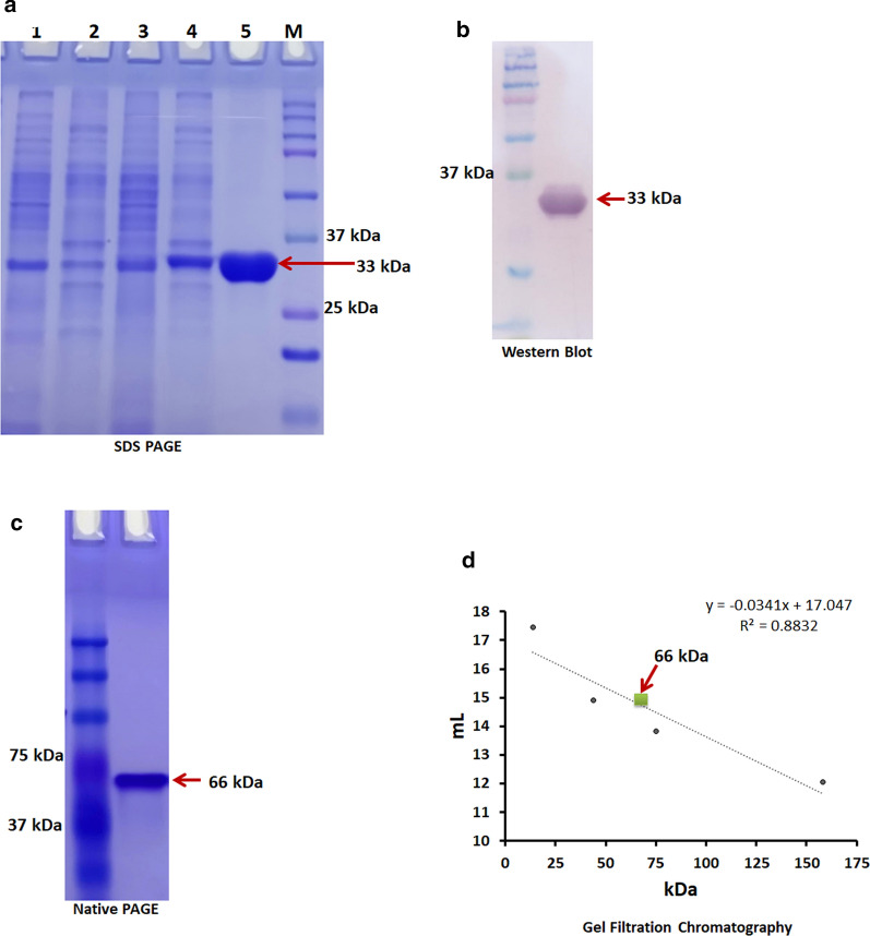
SDS-PAGE analysis of d-allulose 3-epimerase (DaeB). Lane 1: Pellet of B. subtilis transformed with vector; lane 2: Crude cell extract of B. subtilis transformed with vector; Lane 3: Pellet of B. subtilis expressing DaeB; Lane 4: Crude cell extract of B. subtilis expressing DaeB; Lane 5: Purified DaeB protein; Lane M: Protein marker (a). Western blot of DaeB protein using monoclonal anti-polyhistidine and antimouse IgG alkaline phosphatase antibody (b). Native-PAGE analysis of DaeB protein (c). Determination of native molecular mass of DaeB protein by gel filtration. The column was calibrated with standard proteins- Ribonuclease (13.7 kDa), Ovalbumin (44 kDa), Conalbumin (75 kDa) and Aldolase (158 kDa). DaeB is labeled with green square (d)

