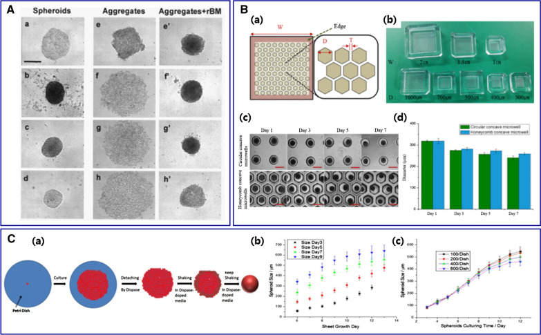
Fig. 4.
A Various morphologies of MCTs depending on cancer cell lines. Compact MCTs were generated with (a) MCF-7, (b) BT-474, (c) T-47D, and (d) MDA-MB-361. (e) MDA-MB-435S cells aggregated tightly but 3 cell lines of (f) MDA-MB-231, (g) MDA-MB-468, and (h) SK-BR-3 aggregated loosely. Adding 2.5% rBM yielded significant compaction (e'–h'). Bar: 500 μm. Reproduced with permission [22].
Copyright 2007, Demetrios Spandidos. B Honeycomb concave microwell. (a) Schematic diagram of a honeycomb concave microwell array (width [W], diameter [D], wall thickness [T]). (b) Various sizes of the honeycomb concave microwell chambers. (c) MCTs formation in the circular and honeycomb concave microwells. Bar: 500 μm. (d) The evaluation of hepatocyte spheroids in 2 different concave microwells [84]. Copyright 2016, Permits unrestricted use. C (a) Illustration of MCTs formation. (b) HCT-116 MCTs size as a function of sheet growth time. The sizes were recorded on different shaking days (days 3, 5, 7, and 9). (c) MCTs size as a function of culturing time with different initial cell seeding density [86]. Copyright 2018, Springer Nature

