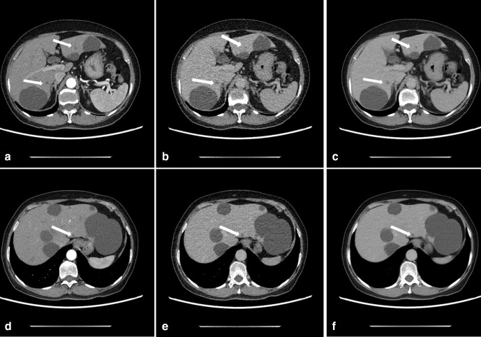
Figure 2.
A 67-year-old female with small hepatic cysts in the arterial-phase (AP) and delayed-phase (DP). CTDIvol value was 6.8 mGy and 1.7 mGy in AP and DP, respectively. A and D: Axial AP images reconstructed with ASIR-V50% shows small hepatic cysts (arrows) with high diagnostic confidence. B and E: Axial DP images reconstructed with ASIR-V50% shows small hepatic cysts (arrows) with low diagnostic confidence due to unclear cyst boundary and high image noise. C and F: Axial DLIR-H DP images for confidence diagnosis of cysts, image noise was significantly reduced, and cyst boundary was clear.

