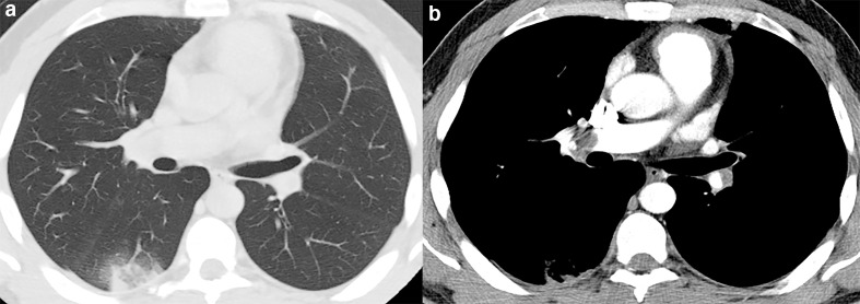
Figure 7.
48-year-old male with pulmonary embolism. (a) Axial CT image demonstrate a wedge-shaped peripheral lung opacity in the right lower lobe with a ground-glass core and a peripheral rim of consolidation yielding the reversed halo sign. (b) CT angiography evidencing the thrombus in the right interlobar artery.

