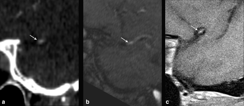
Figure 2.
53-year-old male with multiple vascular risk factors presenting with worsening dementia and multiple infarcts on CT of different stages of evolution (not shown), including right MCA territory acute infarct on MRI (not shown). CTA of the head (a) shows narrowing of the distal right M1 MCA, which measured 39% stenosis (arrow). TOF-MRA (b) showed narrowing at the same location (arrow), with 50% stenosis, while sagittal T 1-weighted IVW (c) showed atherosclerotic plaque along the superior wall of the arterial segment which showed partial enhancement (arrow), resulting in 35% stenosis, which more closely approximated the degree of stenosis seen on CTA relative to MRA.

