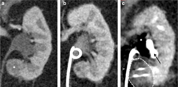
Figure 11.
(A) Coronal contrast-enhanced CT of a 68-year-old female demonstrating the proximity of the medial left lower pole papillary renal cell carcinoma (asterisk) to the ureter. (B) Prior to treatment, a ureteric stent was placed in a retrograde manner. (C) Intraprocedural image demonstrating the ice ball (dashed line) encompassing the lesion and extending right up to the ureter. Carbon dioxide (asterisk) was also injected into the renal sinus to protect the calyces that are opacified and displaced laterally (arrow) and carbon dioxide is also tracking adjacent to the ureter (arrowhead). The stent was removed at 6 weeks post-procedure, with no evidence of a collecting system or ureteric injury.

