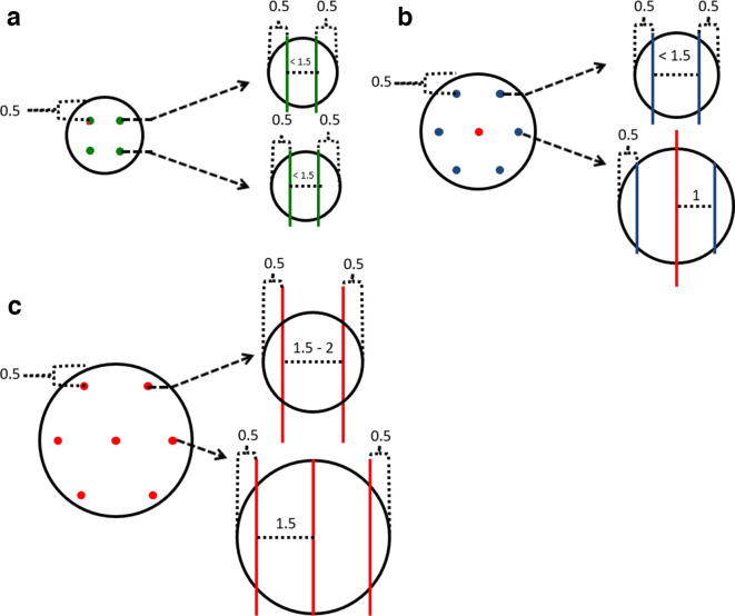
Figure 4.
A guide of where to place Galil cryoprobes and which probes to select for lesions of varying sizes. Images are not to scale and real-life measurements must be made to reflect the imperfect shape of in vivo tumors. Measurements are in cm. The IceSeed, IceSphere and IceRod probes are indicated by green, blue and red colors respectively. Spherical lesions of (A) 2 cm, (B) 3 cm and (C) 4 cm. Coronal images are on the left and the dashed arrows highlight the axial image at that specific level on the right. It is important to stress this is a guide and parallel probe placement at every level is frequently not possible due to anatomical constraints. It is also important to emphasise that the AP positioning of the cryoprobes requires knowledge of the AP length of the isotherm and consideration of whether there are critical structures anterior or posterior to the kidney. Measuring back the length of the expected isotherm along the cryoprobe helps placement in this regard. Some cryoprobes are placed over 1.5 cm from each other in (C) and additional cryoprobes may be required for a central tumour. AP, anteroposterior.

