Figure 5.

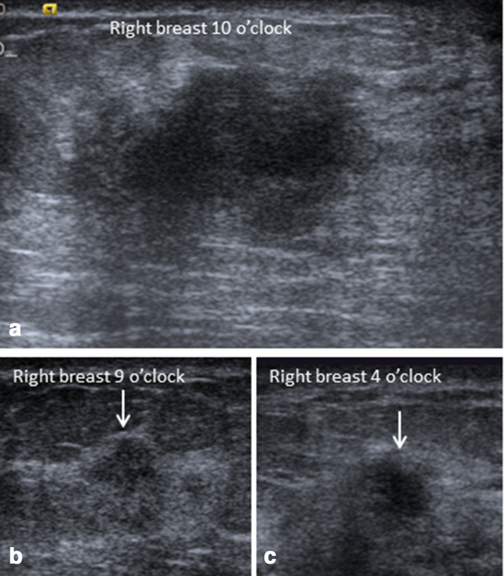
Ultrasonography of the right breast of the same patient in Figures 3 and 4, showed hypoechoic irregular primary mass at 10 o’clock with two additional smaller masses at 9 and 4 o’clock (arrows in b and c).

Ultrasonography of the right breast of the same patient in Figures 3 and 4, showed hypoechoic irregular primary mass at 10 o’clock with two additional smaller masses at 9 and 4 o’clock (arrows in b and c).