
Figure 6.
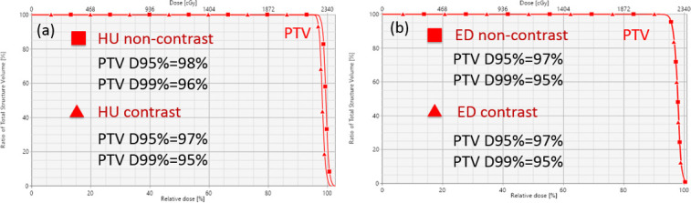
Dose–volume histograms for an example patient (Patient 9) planned for photon therapy. (a) Optimization (non-contrast) vs verification (contrast) for the SECT method. (b) Optimization (non-contrast) vs verification (contrast) for the DLCT method. DLCT, dual layer CT; ED, electron density; HU, Hounsfield unit; PTV, panning target volume; SECT, single-energy CT; SPR, stopping-power ratio.