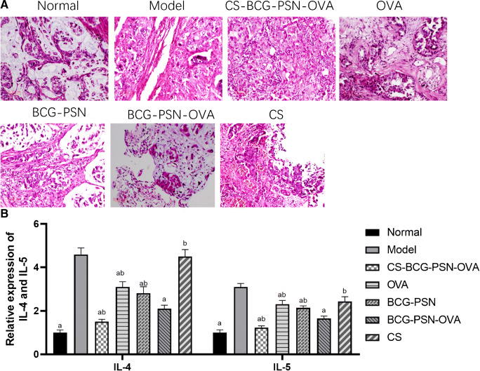
Fig. 5.
Histopathological changes of lung tissue and expression of IL-4 and IL-5 in asthmatic mice after administration. (A) HE staining was used to observe the pathological change of lung tissue in asthmatic mice after administration. (B) The mRNA expression levels of IL-4 and IL-5 in lung tissue cells of asthmatic mice after administration were detected by qRT-PCR. “a” represents a significant difference compared with the Model group (p<0.05). “b” indicates a significant difference compared with the BCG-PSN-OVA group (p<0.05)

