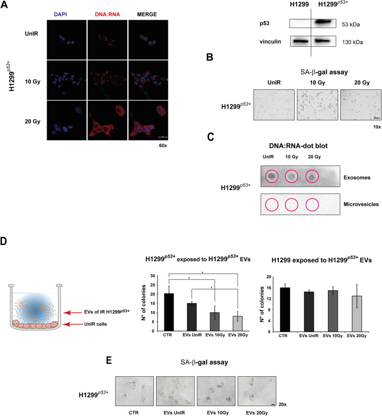
Fig. 5.
H1299 with forced wtp53 (H1299p53+) showed irradiation response similar to that observed in A549 cells. a H1299 cells with forced wtp53 showed a high level of DNA:RNA hybrid structures after irradiation. Cell images were captured by Nikon Eclipse Ti2 confocal microscope with 60x plan apochromat oil immersion objective lens. b. Representative images of senescence-associated β-galactosidase (SA-β-gal) staining detected after H1299p53+ cell exposure to 10 Gy or 20 Gy. Scale bar is 100 μm. c Dot blot analysis of RNA extracted from exosome or microvesicle fraction 72 h post-irradiation and immunoblotted with S9.6 antibody. d Colony-forming assay (CFA). Cells were exposed to EVs isolated from UnIR or IR H1299p53+ cells for 14 days. 0.05% crystal violet solution was used to visualize the generated colonies with more than 50 cells, which were quantified under inverted microscope (Olympus IX51 microscope, Olympus Corporation, Tokyo, Japan) by two independent observers. e Representative images of SA-β-gal staining of UnIR H1299p53+ cells after exposure to EVs isolated from culture medium of IR H1299p53+ cells

