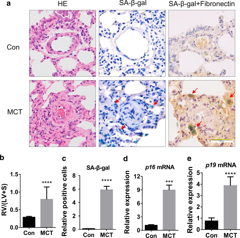
Fig. 1.
Adventitial fibroblasts senescence is observed in the MCT-induced PH rat model. a. Representative images of H&E staining, SA-β-gal staining and Fibronectin-IHC with SA-β-gal staining of rat lung tissues in MCT and control groups. b RV/(LV+S) was determined in each group (n = 8 rats per group). c The quantification of SA-β-gal positive cells normalized to the Con group in Fig. 1a (three pulmonary arteries per rat). d, e. Fold change by qRT-PCR: relative mRNA level of p16/β-actin and p19/β-actin in lung tissues of MCT rats to that in control rats (n = 3 rats per group). All data are shown as the mean ± SEM. Statistical significance compared to controls was assessed using the unpaired two-tailed Student’s t test: ***P < 0.001, ****P < 0.0001

