Fig. 3.
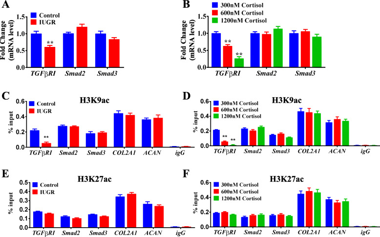
Decreased H3K9ac level of TGFβRI mediated the poor chondrogenic differentiation of WJ-MSCs. a RT-qPCR analysis of TGFβRI, Smad2, and Smad3 after chondrogenic differentiation in the control and IUGR groups. b RT-qPCR analysis of TGFβRI, Smad2, and Smad3 after chondrogenic differentiation in the 300, 600, and 1200 nM cortisol groups. n = 5. c, e ChIP-PCR analysis of H3K9ac and H3K27ac levels of TGFβRI, Smad2, and Smad3, COL2A1 and ACAN after chondrogenic differentiation in the control and IUGR groups. n = 3. d, f ChIP-PCR analysis of H3K9ac and H3K27ac of TGFβRI, Smad2, and Smad3, COL2A1 and ACAN after chondrogenic differentiation in the 300, 600, and 1200 nM cortisol groups. n = 3. H3K9ac, histone 3 lysine 9 acetylation; H3K27ac, histone 3 lysine 27 acetylation; RT-qPCR, real-time quantitative polymerase chain reaction; TGFβRI, transforming growth factor β receptor I; COL2A1, α1 chain of type II collagen; ACAN, aggrecan; WJ-MSCs, Wharton’s jelly-derived mesenchymal stem cells; IUGR, intrauterine growth retardation; ChIP-PCR, chromatin immunoprecipitation-polymerase chain reaction. Data are Mean ± S.E.M. **P < 0.01 vs control

