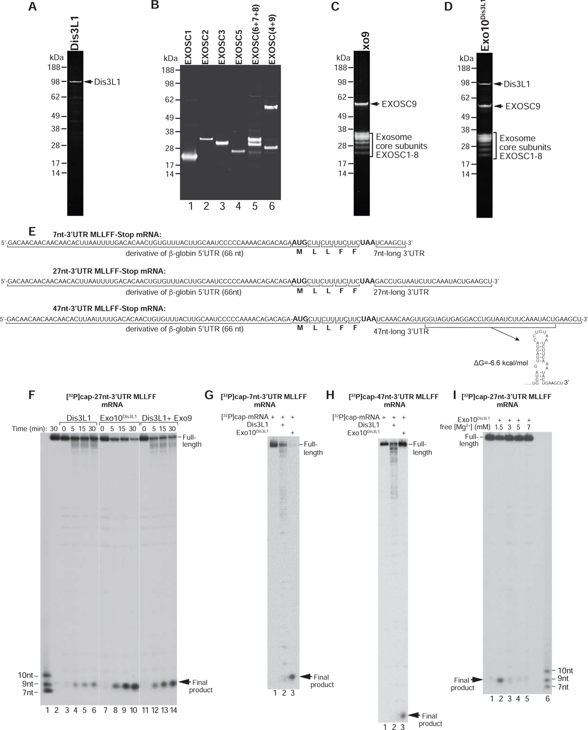
Figure 2. Reconstitution and activity of the mammalian cytoplasmic RNA exosome.

(A) Dis3L1 purified from HEK293T cells, (B) Exo9 subunits purified from E. coli, (C) assembled Exo9 and (D) assembled Exo10Dis3L1, analyzed by SDS-PAGE followed by fluorescent SYPRO staining. (E) Sequences of MLLFF-Stop mRNAs containing 7nt-, 27nt- and 47nt-long 3’UTRs, and the Mfold stem-loop in the 47nt-long 3’UTR. (F) Time courses of degradation of [ 32P]cap-27nt-3’UTR MLLFF-Stop mRNA by Dis3L1, Exo10Dis3L1 and Dis3L1 with Exo9 at 1.5 mM free [Mg2+]. Lane 1 contains RNA markers. Separation of lanes by white lines indicates that they were juxtaposed from the same gel. (G, H) The activities of Dis3L1 and Exo10Dis3L1 on (G) [32P]cap-7nt-3’UTR and (H) [ 32P]cap-47-nt-3’UTR MLLFF-Stop mRNAs after 30 min incubation at 1.5 mM free [Mg2+]. (I) [Mg2+]-dependence of the activity of Exo10Dis3L1 on [32P]cap-27nt-3’UTR MLLFF-Stop mRNA after 30 min incubation. Lane 6 contains RNA markers.
