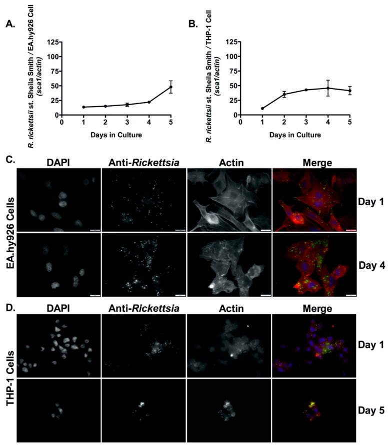
Figure 1.
R. rickettsii st. Sheila Smith proliferates inside both endothelial cells (EA.hy926) and human derived macrophage cells (THP-1). (A,B) EA.hy926 cells and PMA-differentiated THP-1 cells were infected with R. rickettsii st. Sheila Smith (MOI = 2.5), and genomic DNA was extracted at each time point post-infection. Each time point represents the ratio of R. rickettsii st. Sheila Smith sca1 to the host cell actin gene amplified from genomic DNA and determined by quantitative PCR (qPCR). Immunofluorescence microscopy growth analyses in EA.hy926 cells at days 1 and 4 post-infection (C,D) in PMA-differentiated THP-1 cells at days 1 and 5 post-infection demonstrate significant intracellular proliferation. DAPI (blue) was used to visualize host cell nuclei; anti-Rickettsia antibody (RcPFA) followed by Alexa Fluor 488 (green) was utilized to reveal R. rickettsii st. Sheila Smith, and Alexa Fluor 546 Phalloidin (red) was used to indicate the host actin cytoskeleton in C and D. Scale bar = 10 μm. A logistic regression test was used to measure significance (p < 0.05) in growth over time in both mammalian cell lines in A and B.

