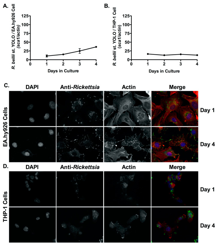
Figure 3.
R. bellii proliferates inside endothelial cells (EA.hy926) but does not grow in human derived macrophage cells (THP-1). (A,B) EA.hy926 cells and PMA-differentiated THP-1 cells were infected with R. bellii st. Yolo (MOI = 2.5), genomic DNA was extracted at indicated time points and growth was determined by qPCR. A logistic regression test was used to measure significance (p < 0.05) in growth over time in both mammalian cell lines. Immunofluorescence microscopy analyses confirmed growth in EA.hy926 cells at days 1 and 4 post-infection (C) but not in PMA-differentiated THP-1 cells at days 1 and 4 post-infection (D). DAPI (blue) was used to stain host cell nuclei; anti-Rickettsia antibody (RcPFA) followed by Alexa Fluor 488 conjugated anti-rabbit IgG (green) was used to stain R. bellii st. Yolo, and AlexaFluor 546-Phalloidin (red) was used to stain actin. Scale bar = 10 μm.

