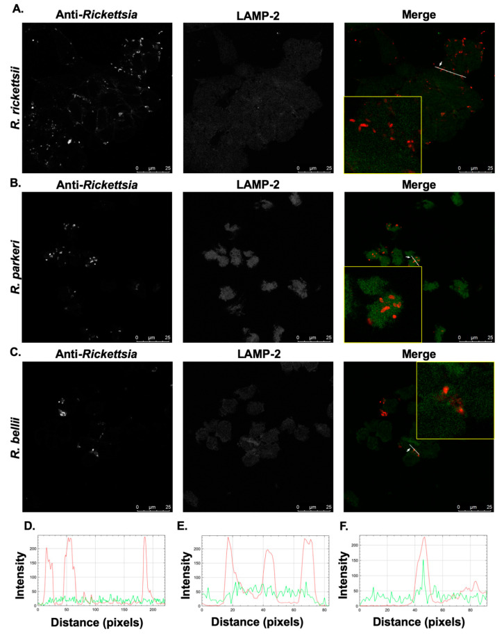
Figure 5.
Pathogenic SFG rickettsiae, but not R. bellii, avoid co-localization with the lysosomal marker, LAMP-2. PMA-differentiated THP-1 cells were infected with (A) R. rickettsii Sheila Smith, (B) R. parkeri Portsmouth, and (C) R. bellii Yolo at MOIs of 10 for 24 h and then processed for immunofluorescence confocal microscopy analyses. Representative slices from z stacks of infected THP-1-derived macrophage 24 h post-infection are shown. (D–F) A generated RGB profile plot documents the relative fluorescence intensity along the indicated white line. Co-localization events were deemed positive when fluorescence intensities from the green and red channels overlap at a given point in the image and negative when intensity peaks do not overlap. Areas of interest for R. rickettsii (D), R. parkeri (E), and R. bellii (F) that were used for determination of co-localization are enlarged to show detail (inset). Scale bar = 25 μm.

