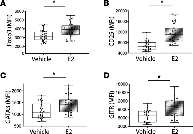
Figure 3. Estrogen enhances the Treg-suppressive phenotype in vitro.
CD4+CD25+ Tregs were isolated from WT mouse splenocytes and cultured in the presence of anti-CD3/CD28 beads and stimulated with either vehicle or estradiol (E2; 10 μM) for 72 hours. Multicolor flow cytometry was performed to asses E2-dependent changes in Treg-suppressive phenotype. Treg expression for Foxp3 (A), CD25 (B), GATA3 (C), and GITR (D) was measured and is expressed as mean fluorescence intensity (MFI) ± SEM. The Mann-Whitney test was used for all MFI. n = 25–30 per group. *P < 0.01.

