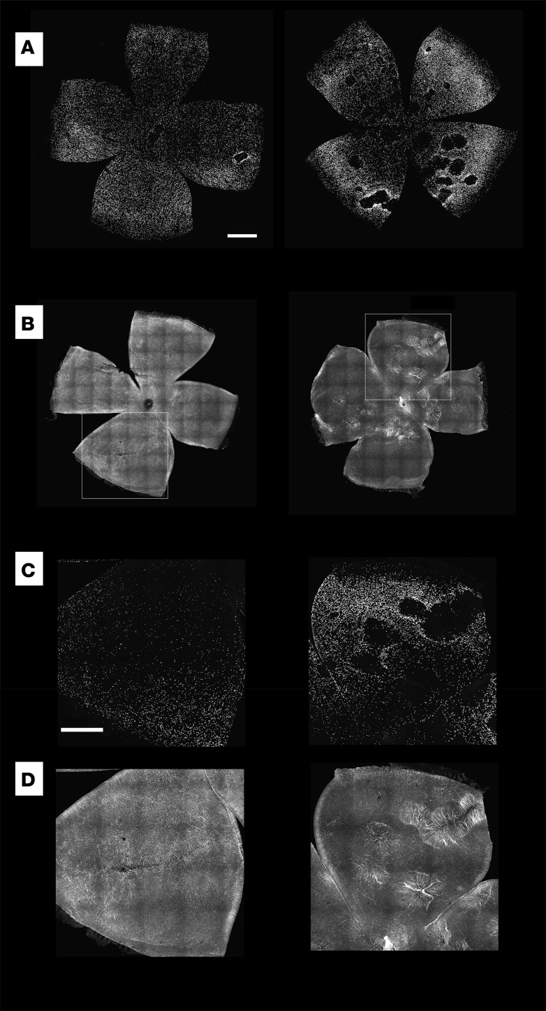
Figure 2. Changes in retinal structure in rd1 mice and the effect of overexpression of Nrf2 in the RPE.
(A) Confocal images of pairs of retina flat mounts from the right and left eyes of a P41 rd1 mice that received a coinjection of AAV-Best1-Nrf2 and AAV-hRedO-H2b-GFP in the left eye (left panel) and control AAV-hRedO-H2b-GFP in the right eye (right panel) (n = 13 mice). Each point of fluorescence is the nucleus of a cone. Note the disruptions, which appear as “holes” or “craters” in the cone mosaic, particularly in the right eyes. (B) Confocal images of a pair of retinas from the right and left eyes of P48 rd1 mouse, with anti-GFAP labeling, demonstrating fairly uniform GFAP distribution in the left retina from the eye that received AAV-Best1-Nrf2 but significant upregulation and distortion of the radial Muller glial fibers in areas of cone loss in the right retina from the eye that received AAV-hRedO-H2b-GFP only (n = 2 mice). Insets show higher magnification of these areas with GFP (C) and GFAP (D). Scale bars: 500 μm for A and B, 200 μm for C and D.

