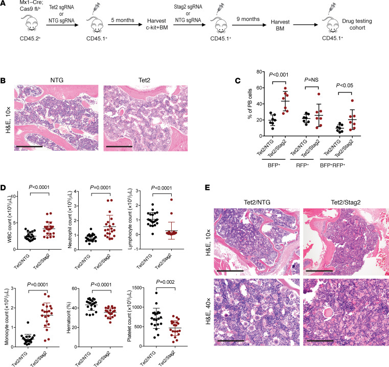
Figure 3. Development of primary models of cohesin-mutant MDS.
(A) Schematic of the sequential bone marrow transplant used to generate Tet2/Stag2-mutant models of myeloid disease. (B) Morphologic evaluation of bone marrow section of mice injected with NTG and Tet2-mutant cells. H&E staining, 10× magnification. No appreciable differences were observed. Scale bar: 0.5 mm. (C) Flow cytometry analysis of peripheral blood (PB) samples of mice sequentially transplanted with Tet2/NTG and Tet2/Stag2 3 months after transplantation. Blue fluorescent protein (BFP) reporter is linked to expression of sgRNA targeting Stag2, and red fluorescent protein (RFP) reporter is linked to expression of sgRNA targeting Tet2. Expansion of BFP+ and BFP+RFP+ cells in Tet2/Stag2 animals. n = 7 per arm. Mean ± SD shown. (D) Absolute white blood cell (WBC) count, neutrophil count, lymphocyte count, monocyte count, hematocrit, and platelet count were measured in Tet2/NTG and Tet2/Stag2-mutant mice 12 weeks after bone marrow transplantation. Mean ± SD is shown. P values were determined using the Student’s t test. n = 20 mice per group. (E) Morphologic evaluation of bone marrow of a representative Tet2/NTG and Tet2/Stag2-mutant mouse shows a decrease in megakaryocytes and increased erythrophagocytosis in Tet2/Stag2-mutant mice. Images were stained using H&E and imaged at 10× (scale bar: 0.5 mm) and 40× (scale bar: 0.125 mm) original magnification.

