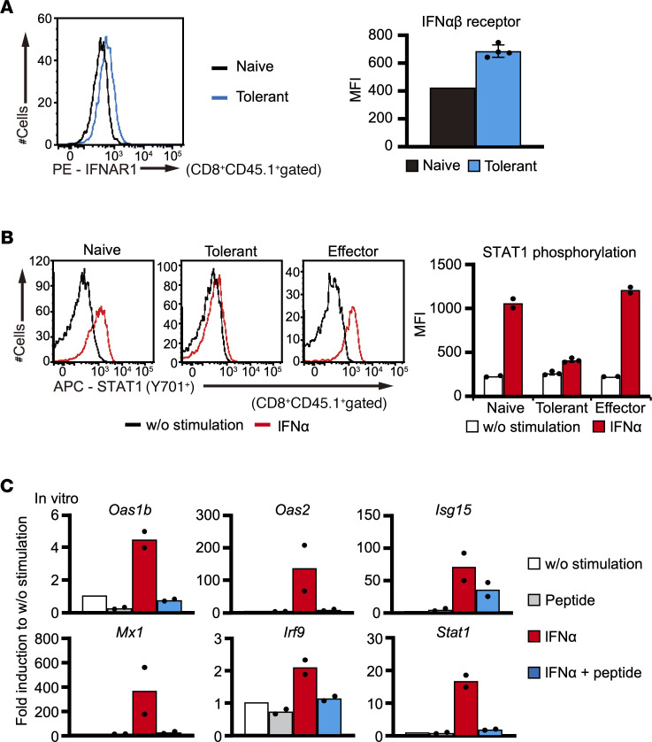
Figure 3. Responsiveness to IFN-α is suppressed in tolerant CD8+ T cells.
(A) IFN-αβ receptor expression in naive (n = 1) and tolerant (n = 4) COR93-specific T cells was analyzed by FACS. (B) STAT1 phosphorylation was examined in naive (n = 2), tolerant (n = 3), and effector (n = 2) COR93-specific T cells after in vitro stimulation with IFN-α and COR93 peptide for 15 minutes. A representative result in each group is shown by histograms (left panels), while individual value and the average of mean fluorescence intensity in each group are shown by a dot plot and a bar graph, respectively (right panels). (C) The expression of IFN-I–related genes in COR93-specific CD8+ T cells were analyzed by RT-qPCR after stimulating spleen cells from COR93-specific TCR-Tg mice with IFN-α and/or COR93 peptide for 8 hours (n = 2 for each group). Data represent mean ± SD (n ≥ 3).

