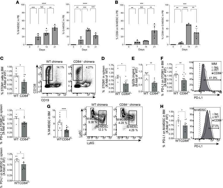
Figure 10. M-MDSC expansion and suppression in vivo is dependent on CD84.
(A and B) 5TGM1 cells were i.v. injected into syngeneic immunocompetent C57BL/KaLwRij mice. PB samples were collected and analyzed after 1 week, 2 weeks, and 3 weeks by weekly submandibular bleeding after the injection. The percentages of M-MDSCs and G-MDSCs (A), and CD84 expression on M-MDSCs and G-MDSCs (B), were analyzed by flow cytometry (n = 5, **P < 0.01, ***P < 0.001, ****P < 0.0001). (C–H) WT or CD84–/– mice were irradiated with 1050 Rad and injected with 1 × 10⁶ C57BL/KaLwRij BM cells after 1 day. After 60 days, the mice were injected with 1 × 10⁶ 5TGM1 cells. After an additional 21 days, the mice were sacrificed, and their BM, blood, and spleens were analyzed. (C and D) Percent of MM cells in the BM (C) and spleen (D). (E) IgG2b antibodies in the blood. (F) PD-L1 protein levels on the surface of MM from BM and spleen. Representative histogram of the BM is shown (n = 9–14, *P < 0.05, **P < 0.01). (G) Percent of M-MDSCs in the BM. Representative dot plots shown (n = 14, ****P < 0.0001). (H) PD-L1 on the surface of M-MDSCs from BM (left graph) and spleen (right graph). Representative histogram of BM is shown (n = 6–10, *P < 0.05).

