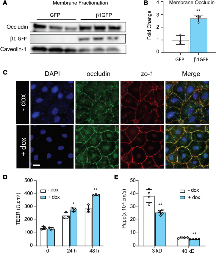
Figure 2. Overexpression of the β1 subunit increases alveolar type I barrier function.
(A) Overexpression of the β1 subunit in HEK293T cells increased occludin expression at the plasma membrane. Caveolin-1 was used as membrane loading control. (B) Densitometry of gels in A, with analysis by Student’s t test, **P < 0.01. (C) ATII cells were cotransfected with 4 mg/mL pCMV-Tet3G plasmid and 16 mg/mL pTet3G-human β1 plasmid day 1 after isolation. Cells were then plated on fibronectin-coated coverslips. Doxycycline (1 μg/mL) was added 48 hours later. Representative immunofluorescence staining of ATI cells shows that doxycycline-induced expression of the β1 subunit induces more mature tight junctions, as indicated by occludin (green) and zo-1 (red) staining. Images represent 3 independent experiments. Scale bar: 20 mm. (D) ATII cells were cotransfected as in C, but cells were plated on fibronectin-coated 12-well transwell plates. Twenty-four hours later at day 2, 1 μg/mL of doxycycline (dox) was added to induce β1 gene expression. TEER was measured every 24 hours. ANOVA followed by Bonferroni’s post hoc test was used for statistical analysis, *P < 0.05, **P < 0.01. (E) After TEER measurement at day 4, permeability to 3 kD Texas Red–dextran and 40 kD FITC-dextran was measured for a duration of 2 hours. Data are presented as mean ± SD. ANOVA followed by Bonferroni’s post hoc test was used for statistical analysis, **P < 0.01.

