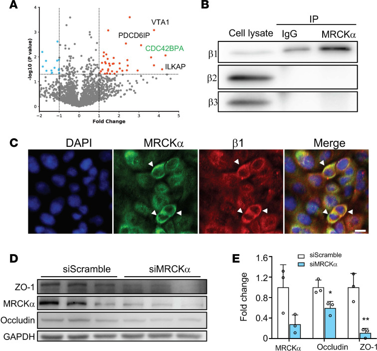
Figure 3. MRCKα interacts with the β1 subunit and stabilizes tight junction.
(A) Volcano plot of proteins identified from triplicate mass spectrometry experiments. CDC42BPA (MRCKα) is labeled on the graph. Dashed line indicates the P value threshold of 0.05. (B) The interaction of MRCKα with the β1 subunit was confirmed using co-IP. A total of 5% of total cell lysate was used for input. The β2 or β3 subunit did not coimmunoprecipitate with MRCKα. (C) The β1 subunit (red) and MRCKα (green) colocalize in ATI cells. Scale bar: 20 μm. (D) ATI cells were transfected with a scrambled siRNA (siScramble) or a siRNA against MRCKα (siMRCKα). (E) Twenty-four hours later, cells were lysed for immunoblot analysis and quantified. Data represent 3 biological replicates and Error bars show SD. Student’s t test, *P < 0.05, **P < 0.01.

