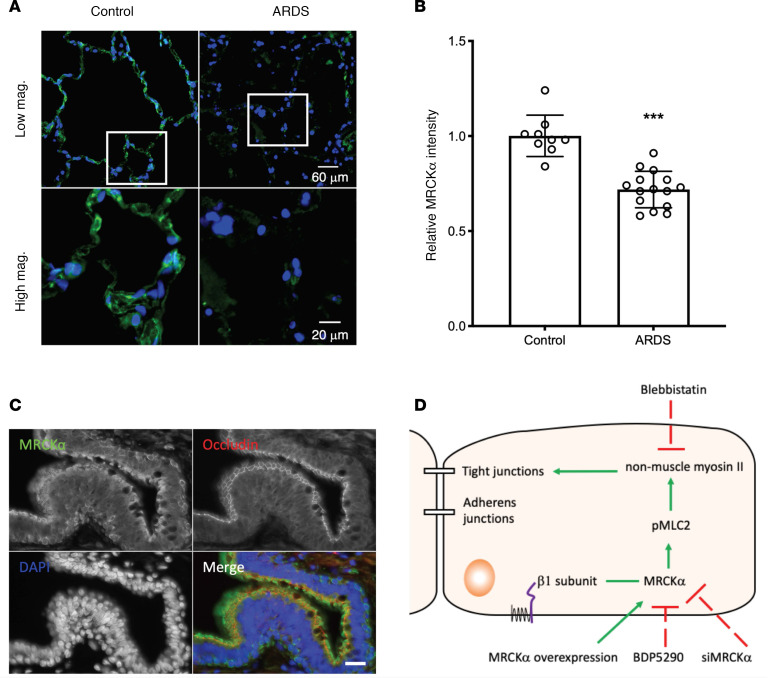
Figure 6. Decreased MRCKα levels in the alveolar epithelium of human ARDS patients.
(A) Representative images of immunofluorescence staining for MRCKα (green) in lung sections of a control donor and a patient with ARDS. Upper panel shows images taken at 20× objective magnification, and lower panel shows images taken at 63× objective magnification for the boxed region in the upper panel. (B) Quantification of MRCKα expression in the alveoli. ROI (region of interest) were drawn in the alveoli region, and the ratio of integrated pixel intensity for MRCKα and DAPI was calculated for each ROI. Three normal donors and 5 ARDS patients were used for quantification, with 3 random fields chosen for each sample. Data are expressed as mean ± SEM, with n = 9 (3 patients) for normal control and n = 15 (5 patients) for ARDS. Statistical analysis was by 2-tailed Student’s t test, ***P < 0.001. (C) Costaining of MRCKα (green) and occludin (red) in the small airway from control donor. Scale bar: 20 µm. (D) Working model of the β1 subunit increases alveolar epithelial barrier integrity. The β1 subunit of the NKA interacts with MRCKα, assists in its activation, leads to higher myosin phosphorylation, and eventually stabilize tight junctions.

