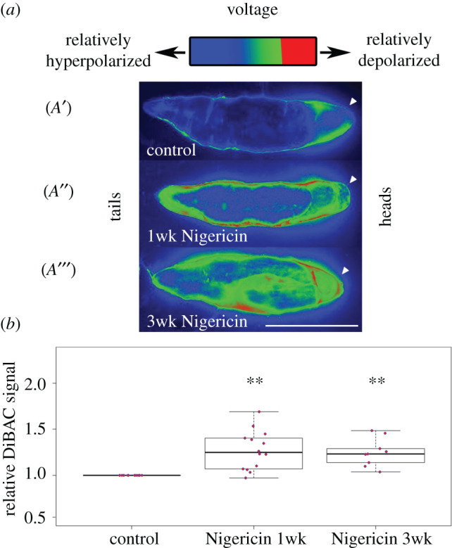
Figure 4.

Bioelectrical long-term memory in planaria. Voltage-sensitive fluorescent dye reveals the spatial distribution of bioelectric states in intact planaria [33]. Our model of the bioelectric pattern as a memory predicts that after editing this pattern, it should remain altered for long periods of time (relative to the normal rate of change of bioelectric parameters, which is milliseconds for the CNS and minutes–hours in developmental bioelectricity). Control animals show depolarization at the anterior end (A′). After soaking in ionophore, which induces a shift to Cryptic phenotype, both ends become relatively depolarized and stay that way for one week (A″). Remarkably, even three weeks later, this altered pattern persists and indeed becomes stronger, as discussed above in the context of consolidation and construction of memories over time (A′′′). Quantification of the altered bioelectric states shows significant differences persisting at one and three weeks after exposure to ionophore.
