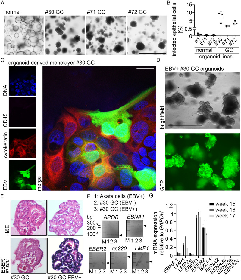
Fig 4. EBV can infect gastric cancer organoids.
(A) Brightfield microscopy of normal and cancer organoids. #1–72 refers to patient IDs. Scale: 1000 μm. (B) At 6 dpi, EBV transfer-infected organoid-derived monolayers (normal and GC) were analyzed for EBV infection rate by flow cytometry. Results are shown as means of three independent experiments with SD. (C) At 4–6 dpi immunofluorescence was performed on EBV transfer-infected organoid-derived monolayers for epithelial marker Cytokeratin, GFP-expressing EBV and lymphocyte marker CD45. DNA was counterstained with Hoechst. Scale: 25 μm. Representative images of three independent experiments. (D) EBV transfer-infected #30 cancer organoid cells were FACS-sorted, clonally expanded and monitored by fluorescence microscopy. Scale: 1000 μm. (E) EBER in situ hybridization, detecting small non-coding RNA of EBV was performed on embedded clonal EBV+ or EBV- cancer organoids. (F) PCR analysis for the presence of EBV DNA (EBER2, EBNA1, gp220 and LMP1) in clonal EBV+ or EBV- cancer organoids. APOB was used as eukaryotic control gene. (G) RT-qPCR was performed on RNA extracted from the infected #30 cancer organoid line. The viral gene expression profile included expression of EBNA1, LMP1 and LMP2a plus the non-coding EBERs.

