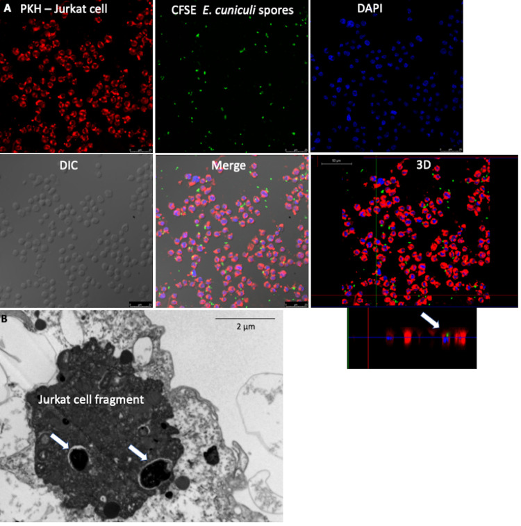
Fig 3. Infection of Jurkat cells (ACs) by E. cuniculi before inducing apoptosis.
(A) Jurkat cells (ACs) stained with PKH; E. cuniculi spores marked with CFSE; core fluorescence stained with DAPI; phase-contrast cultures-DIC; overlay of images showing internalized and intact spores inside the Jurkat cells, confirmed in the 3D image (arrow). (Images) (B) Ultramicrographic images of a Jurkat cell fragment infected with E. cuniculi (arrow).

