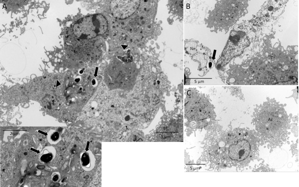
Fig 6. Ultramicrography image of macrophages pre-incubated with E. cuniculi in infected and apoptotic cells.
(A) Apoptotic bodies phagocyted (arrowhead) by macrophages containing intact E. cuniculi spores inside (insert) (B) necrotic cells (Ne) with the spores of pathogen close to macrophages. (C) A macrophage in apoptosis (Ac).

