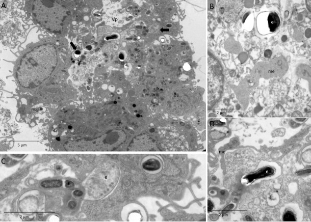
Fig 7. Ultramicrography image of pre-incubation of macrophages with infected and apoptotic Jurkat cells after challenge with E. cuniculi.
(A) Parasitophore vacuole (Vp) with E. cuniculi spores (arrows) and other forms of development. (B) Detail of meronte (me) and spores (e) with rupture of resin, characteristic of microsporidia. (C) Detail of immature spores (e) showing an enameled polar tubule. (D) Detail of spores (e) with rupture of resin, characteristic of microsporidia and meronte (me) next to the Vp membrane.

