
Fig. 1. BF cholinergic neurons provide robust inputs onto top-down ACAVIS projection neurons.
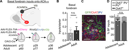
(A) Experimental overview of rabies virus–mediated input mapping of BF inputs onto ACAVIS neurons in adolescent and adult mice. (B) Cre-dependent AAVs encoding TVA-mCherry and Rb-G were injected into ACA and retrograde CAV2-Cre into VIS. After 2.5 weeks, pseudotyped rabies virus was injected into ACA to allow for uptake of rabies virus in ACAVIS neurons to express GFP in input cells over 1 week. AAV injections were initiated at p12 to capture inputs during adolescence (p36) and at p60 to capture inputs during adulthood (p84). (B) Number of BF inputs, when normalized to the number of mCherry + eGFP + starter cells in ACA (t test, t11 = 0.5241, P = 0.611; adolescent: seven mice; adult: six mice). n.s., not significant. (C) Representative image of immunohistochemical characterization of BF GFP + inputs by choline acetyltransferase (ChAT; red) and parvalbumin (PV; blue). Scale bar, 50 μm. LPO, lateral preoptic nucleus; NDB, nucleus of the diagonal band. (D) Inputs from BF are primarily ChAT+ during adolescence (82.5%) and adulthood (78.17%), with a smaller PV+ population (adolescent, 6.775%; adult, 4.467%), and ChAT-PV− (adolescent, 10.73%; adult, 17.37%). The relative proportion of cell types does not differ between adolescent and adult groups (t test, t5 = 0.4904, P = 0.6446; adolescent: four mice; adult: three mice).