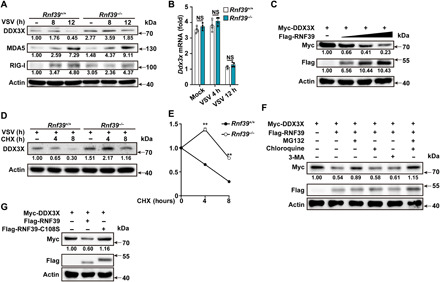
Fig. 6. RNF39 promotes proteasomal degradation of DDX3X.

(A and B) Immunoblot analysis of extracts (A) or RT-PCR analysis (B) of mouse PMs from Rnf39+/+ or Rnf39−/− mice infected with VSV for the indicated time points. (C) Immunoblot analysis of extracts from HEK293T cells transfected with Myc-DDX3X and increasing amount of Flag-RNF39 (0, 0.5, 1, or 2 μg) expression plasmids. (D and E) Immunoblot analysis of extracts (D) from Rnf39+/+ or Rnf39−/− mouse PMs infected with VSV for 4 hours and then treated with cycloheximide (CHX) for various times. DDX3X expression level was quantitated by measuring band intensities using “ImageJ” software (E). The values were normalized to actin. (F) Immunoblot analysis of extracts from HEK293T cells transfected with Myc-DDX3X and Flag-RNF39 expression plasmid and then treated with MG132, chloroquine, or 3-MA for 4 hours. (G) Immunoblot analysis of extracts from HEK293T cells transfected with Myc-DDX3X, together with Flag-tagged RNF39 or RNF39 C108S expression plasmids. All data are represented as means ± SD. Significance was determined by unpaired two-tailed Student’s t test: **P < 0.01. All experiments were repeated at a minimum of three times.
