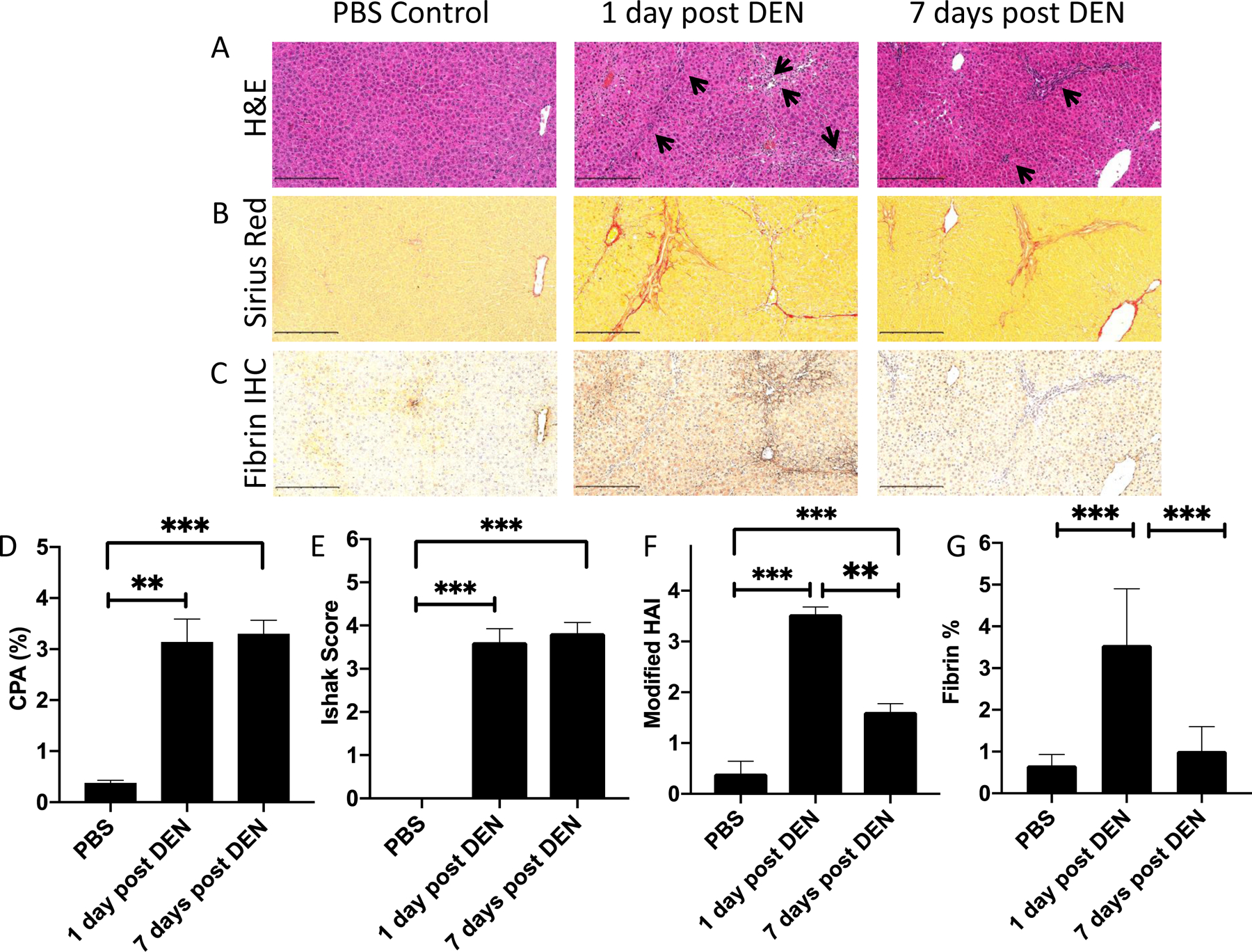
Figure 1.

Histological analysis of liver specimens from rats given 4 weekly i.p. doses of PBS (left) or 100 mg/kg of DEN and sacrificed at 1 day (middle) or 7 days (right) following the last DEN administration, scale bar = 250 μm. (A) Hematoxylin and eosin (H&E) staining of liver tissue. Inflammatory regions are labeled with pointed black arrows. (B) Sirius Red staining of liver tissue. Fibrosis is shown by intense red staining of collagen fibers. (C) Immunohistochemistry staining of fibrin in liver tissue. (D) Morphometric analysis of Sirius Red stained slides to give collagen proportional area showing moderate fibrosis in both DEN groups that is significantly higher than in PBS controls. (E) Ishak scoring of liver fibrosis showing moderate fibrosis in both DEN groups that is significantly higher than in PBS controls. (F) Histological activity index for inflammation (0 – 4) showing that inflammation is significantly higher at 1 day post DEN than 7 days post DEN. (G) Morphometric analysis of the area of the slide stained positive for fibrin by immunohistochemistry. ** P<0.01, *** P<0.0001, one way ANOVA with post hoc Tukey test.
