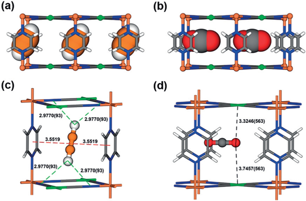
Figure 4.
Neutron diffraction crystal structure of a) FeNi-M’MOF⊃C2D2 and b) FeNi-M’MOF⊃CO2, viewed from the a/b axis. Adsorption binding sites of c) C2D2 and c) CO2 for FeNi-M’MOF. Fe, Ni, C, N, O, H in FeNi-M’MOF and CO2 are represented by orange, green, gray, blue, red, and white, respectively; C and D in C2D2 are represented by orange and white, respectively. The labelled distance is measured in Å.

