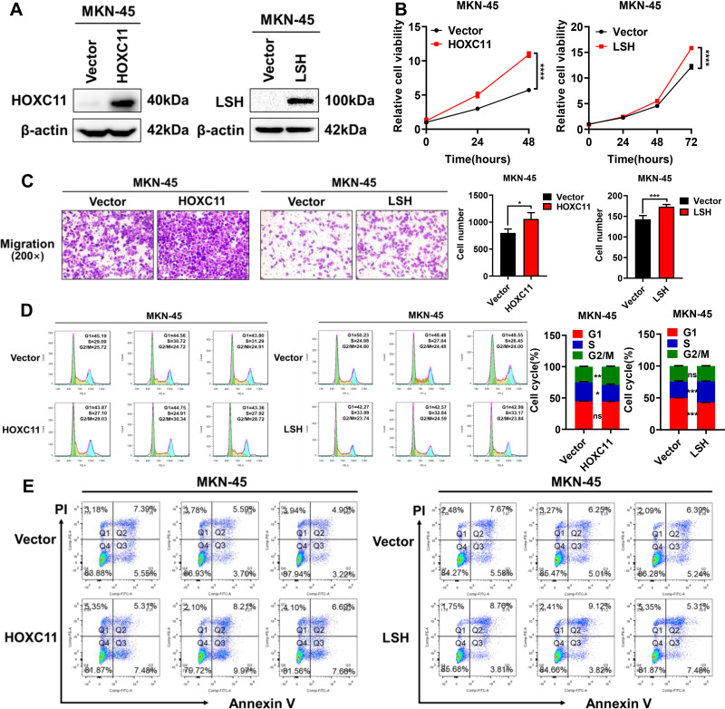
Figure 5.
Overexpression of HOXC11 and LSH promotes GAC progression. (A) The establishment of HOXC11 and LSH over-expressed cell line detected by Western blot. (B) Cell viability was detected by CCK-8. Shown is the mean ± SD of experiments (n=5), ****, P < 0.0001. (C) HOXC11 and LSH overexpression enhanced migration of MKN-45 cells. Shown is the mean ± SD of experiments (n=3), *P < 0.05, ***P < 0.001. (D) HOXC11 and LSH accelerates cell cycle detected by flow cytometry. Shown is the mean ± SD of experiments (n=3), *P < 0.05, **P < 0.01, ***P < 0.001, ns, not significant. (E) Flow cytometry detected the cell apoptosis of HOXC11 and LSH.

