. 2021 Feb 22;10:e63819. doi: 10.7554/eLife.63819

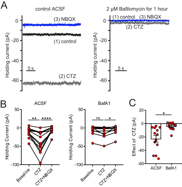
Figure 6. Depletion of glutamate from synaptic vesicles confirms vesicular source of ambient glutamate.

(A) Example recordings of the baseline current of an ON UBC in control (left panel) and of an ON UBC after BafA1 treatment (right), showing the decrease in the current revealed by 100 µM CTZ after treatment with BafA1, with subsequent block by 5 µM NBQX. (B) Left: After incubation with ACSF as a control, CTZ increased the inward holding current significantly, which was reversed by NBQX. Friedman test, p<0.0001, n = 12; Dunn's multiple comparisons tests, baseline vs CTZ, p=0.01, CTZ vs CTZ+NBQX, p<0.001. Right- Incubation in BafA1 reduced the effect of CTZ. Friedman test, p=0.0169, n = 12; Dunn's multiple comparisons tests, Baseline vs CTZ, p=0.459; CTZ vs CTZ+NBQX, p=0.0128. (C) The effect of CTZ was significantly reduced by incubation in BafA1, unpaired t-test, p=0.002, n = 25.

Figure 6.

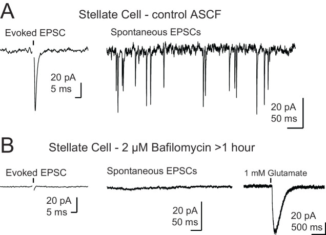
Figure 6—figure supplement 1. Control for recordings in the presence of bafilomycin A1.

Figure 6—figure supplement 1.

(A) Example traces of a control stellate cell with evoked (left) and spontaneous (right) EPSCs. (B) Example traces from a stellate cell after BafA1 treatment and reduction of vesicular glutamate concentration. Evoked EPSCs and spontaneous EPSCs were absent. Far right trace shows a reliable response to puff application of glutamate demonstrating that postsynaptic receptors were present and functioning. Stellate cells (n = 3) were filled with Alexa 488, and the dendrite locations were easily identified by fluorescence for targeted puff. Black arrows in (A) and (B) show onset of a single electrical stimulus and in (B) black arrowhead indicates onset of a brief puff (10–15 ms) of glutamate. Stimulus artifact was removed for clarity.