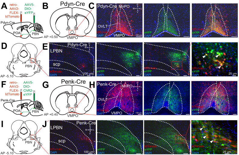
Figure 3. Pdyn+ and Penk+ LPBN neurons project to VMPO.
(A) Illustration of injection of retroAAV-DIO-tdTomato in POA and AAV5-DIO-eYFP in a Pdyn-Cre mouse. (B) Diagram of POA region depicted in (C) showing antero- (green) and retrograde (red) labeling of Pdyn+ neurons in POA. (D) Diagram of parabrachial nucleus (PBN) region depicted in (E) showing retrograde labeling from POA (red) and eYFP expression (green). Yellow cells in overlay image, marked with arrow heads, illustrate dual labeling by locally injected and retrograde viruses. (F) Illustration of injection of retroAAV-DIO-tdTomato in POA and AAV5-DIO-eYFP in an Penk-Cre mouse. (G) Diagram of POA region depicted in (H) show antero- (green) and retrograde (red) labeling of Penk+ neurons in POA. (I) Diagram of PBN region depicted in (J) showing retrograde labeling from POA (red) and eYFP expression (green). Yellow cells in overlay image, marked with arrow heads, illustrate dual labeling by locally injected and retrograde viruses.

