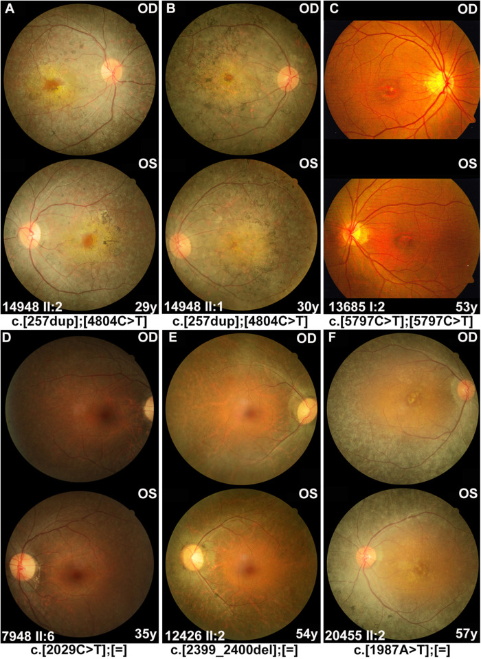
FIGURE 2.
Fundus photographs of the affected individuals with RP1 variants. (A,B) Severe phenotypes were shown in patients with biallelic RP1 variants, including waxy pale optic disc, attenuated vessels, and periphery degeneration with bone spicule pigmentation and involving macular region. (C) A 53 year-old patient diagnosed with macular degeneration rather than RP, who carried homozygous variant c.5797C > T (p.Arg1933*) in our cohort. (D–F) Patients with heterozygous RP1 variants were characterized by peripheral pigment disorders and scattered distribution of bone spicule pigmentation, both of which were often absent in the macular region.

