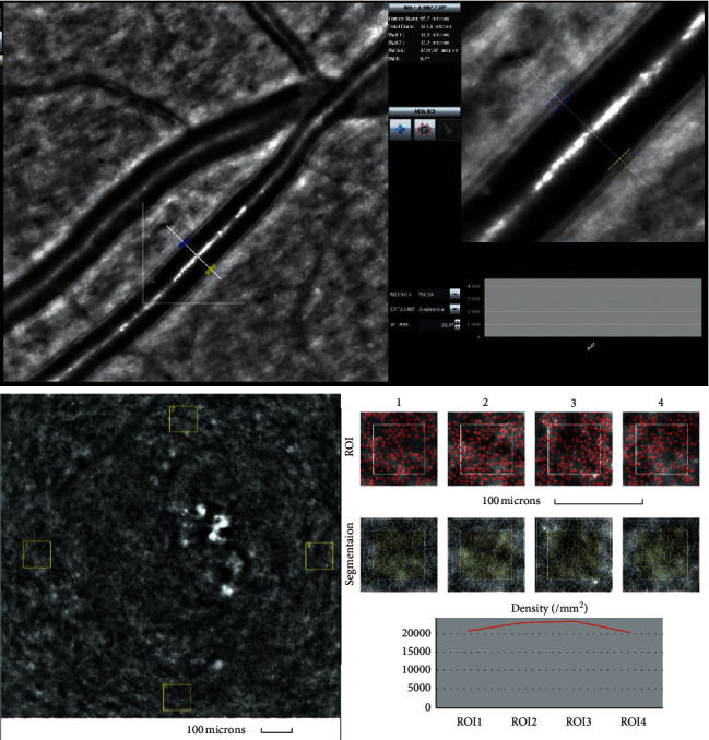
Figure 2.

Image of the retinal artery (upper) and of four retinal cones squares windows (cones depicted in red and Voronoi triangulation) (lower) for a patient from the control group (IK) captured by rtx1 AO retinal camera. The following parameters are shown: in the artery analysis; Lumen: 96.7 μm, Total diameter: 121.4 μm, Wall_1: 12.5 μm, Wall_2: 13.2 μm, WLR- 0.27, WCSA- 4277.0 μm2, AL- 22.87 mm; in the cone analysis: for different ROI (region of interest) squares: Regularity: 82.9–92.5%, Cone spacing: 7.21–7.71 μm, Cone density: 20264–22831 cone/mm2.
