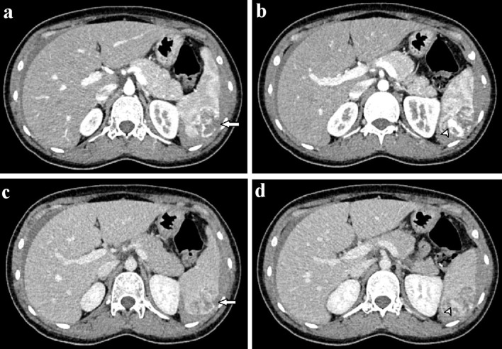
Figure 2.
Axial abdominal CT in the arterial (a, b) and venous phases (c, d). A 3 × 4 cm mass with ill-defined borders was observed in the spleen (a). This mass showed heterogeneous enhancement and hypervascularity (a, b). The mass enhanced heterogeneously on the venous phase (c). A tumor laceration was observed on the splenic capsule (a, c), resulting in intratumor pseudoaneurysm (b, d; arrowhead). CT: computed tomography.

