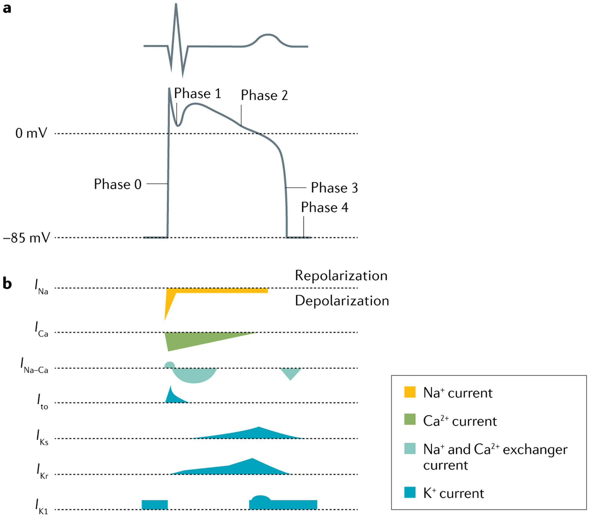
Fig. 3 |. Ventricular action potential and ionic currents.

a | Schematic showing a normal electrocardiogram trace and the corresponding phases of the ventricular action potential (0, 1, 2, 3 and 4) that determine the shape of the trace. b | The major transmembrane ionic currents that generate the ventricular action potential. Inward currents that contribute to depolarization are oriented downwards, and outward currents that contribute to repolarization are oriented upwards; the shapes of the currents indicate their relative intensity. Ito, transient outward potassium current.
