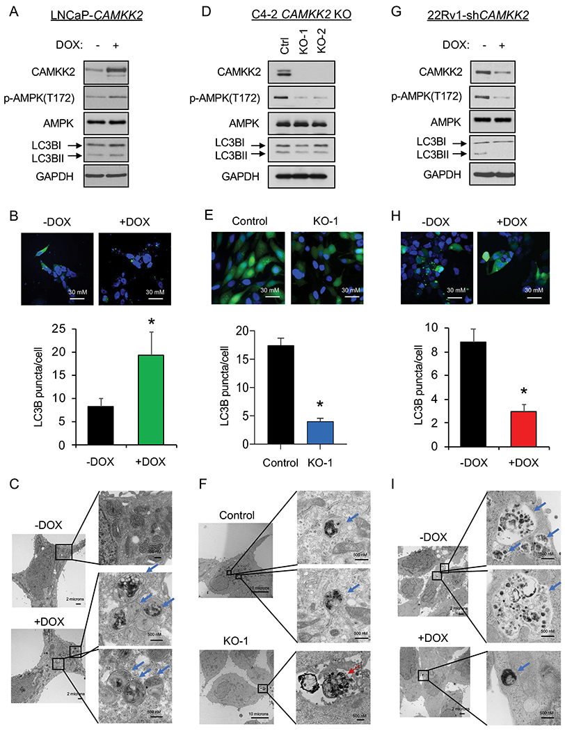
Fig 2. CAMKK2 increases autophagy in prostate cancer cells.

(A) Immunoblot analysis of doxycycline (DOX)-inducible LNCaP stable cells (LNCaP-CAMKK2) that express CAMKK2 upon addition of 50 ng/ml DOX for 48 hours. (B) LNCaP-CAMKK2 cells were transiently transfected with GFP-LC3 (green) and then treated ± 50 ng/ml DOX for 48 hours. Representative images (top). GFP-LC3 puncta (green) were quantified as the average number of GFP-LC3 puncta per cell ± SEM (bottom). The nuclei are stained with DAPI (blue) for reference. P value was calculated using a two-tailed t test. *P < 0.05. (C) LNCaP-CAMKK2 cells were treated ± 50 ng/ml DOX for 48 hours and imaged using transmission electron microscopy (TEM). Two magnifications of ultrastructures are shown. Blue arrows indicate autophagosomes and autolysosomes. (D) Immunoblot analysis of two independent clones of CRISPR-modified C4-2 CAMKK2 knockout (KO) cells compared with their parental C4-2 Cas9 control cells (Ctrl). (E) GFP-LC3 was expressed in C4-2 Cas9 control and CAMKK2 KO cell derivatives. GFP-LC3 puncta (representative images; top) and quantification (bottom) are shown as in B. (F) C4-2 control and C4-2 CAMKK2 KO cells were imaged using TEM as in C. Red arrows indicate apoptotic bodies. (G) Immunoblot analysis of DOX-inducible 22Rv1 stable cells that express shRNA targeting CAMKK2 (22Rv1-shCAMKK2) with 800 ng/ml DOX treatment for 72 hours. (H) 22Rv1-shCAMKK2 cells were transiently transfected with GFP-LC3 and then treated ± 800 ng/ml DOX for 72 hours. GFP-LC3 puncta (representative images; top) and quantification (bottom) are shown as in B. (I) 22Rv1-shCAMKK2 cells were treated ± 800 ng/ml DOX for 72 hours and imaged with TEM as in C.
