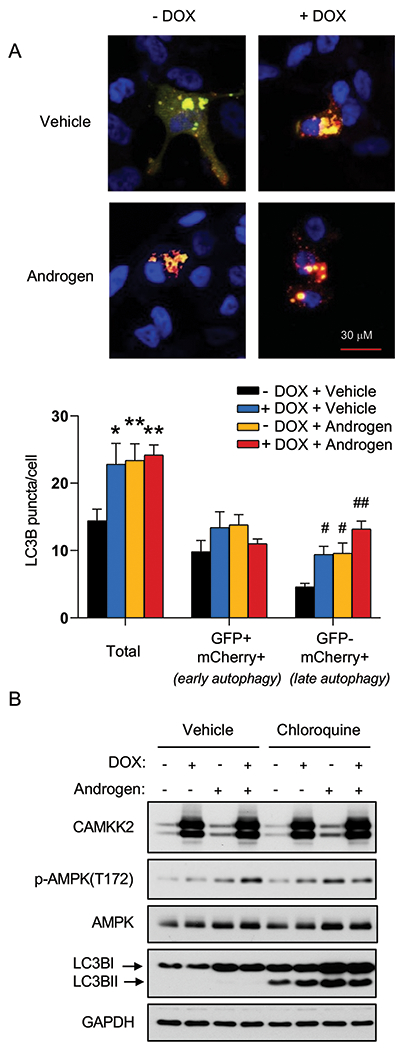
Fig 3. CAMKK2 promotes autophagic flux.

(A) LNCaP-CAMKK2 cells were transfected with an mCherry-GFP-LC3 plasmid and treated ± 10 nM R1881 (androgen) ± 50 ng/ml DOX. Representative fluorescence images of the cellular localization of autophagic puncta (top) and quantification (bottom). P values were calculated using one-way ANOVA with Dunnett’s test. *P < 0.05, **P < 0.01, compared to vehicle group in total. #P < 0.05, ##P < 0.01, compared to vehicle group in GFP-mCherry+. (B) LNCaP-CAMKK2 cells were treated ± 10 nM R1881 (androgen) ± 50 ng/ml DOX ± 20 μM chloroquine (lysosomal block/inhibitor of autophagic flux) for 72 hours. Cell lysates were then subjected to immunoblot analysis.
