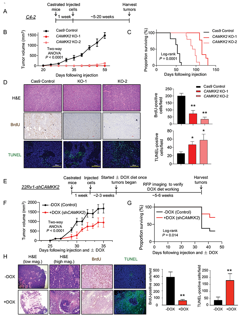
Fig 4. CAMKK2 is required for CRPC tumor growth in vivo.

(A) Schematic of xenograft study using CRPC C4-2 Cas9 control and CAMKK2 CRISPR knockout (KO) cell derivatives in castrated NSG mice. (B) Tumor growth curves of C4-2 Cas9 control and C4-2 CAMKK2 KO xenografts in castrated NSG mice (n = 10/group). P values were calculated using two-way ANOVA. (C) Kaplan-Meier survival curve of C4-2 Cas9 control and C4-2 CAMKK2 KO xenograft mice. P values were calculated using the log-rank test. (D) C4-2 xenograft tumor samples were stained with H&E, BrdU and TUNEL. Representative images (left) and quantifications of BrdU and TUNEL staining (right). *P < 0.05, **P < 0.01 by one-way ANOVA with Dunnett’s test. (E) Schematic of xenograft study using DOX-inducible CRPC 22Rv1-shCAMKK2 cells in castrated NSG mice. (F) Tumor growth curves of 22Rv1-shCAMKK2 xenografts in castrated NSG mice fed control or DOX-enriched (625 mg/kg) chow. P value was calculated using two-way ANOVA. (G) Kaplan-Meier survival curve of 22Rv1-shCAMKK2 xenograft mice ± DOX. P value was calculated using the log-rank test. (H) 22Rv1-shCAMKK2 xenograft tumor samples were stained with H&E, BrdU and TUNEL. Representative images (left) and quantifications of BrdU and TUNEL staining (right). Note, evidence of perivascular tumor sparing in DOX-treated tumors (H&E high magnification (mag.)). **P < 0.01 by t test.
