FIGURE 2.

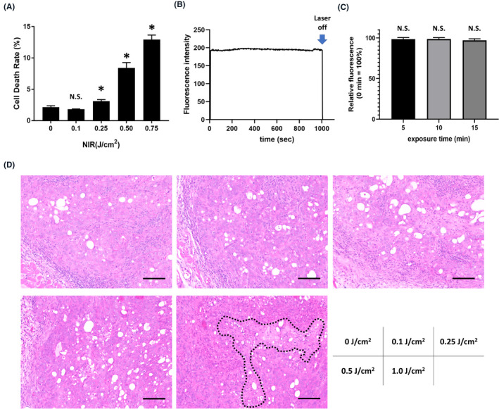
Therapeutic effect and fluorescence decay by weak near‐infrared (NIR) laser. A, Cell viability after weak NIR laser irradiation (1 mW/cm2). Cell viability was measured by propidium iodide (PI) staining. Data are presented as mean ± SEM (n = 4, *P < .05 vs no light exposure group). B, Time course of tumor fluorescence intensity during weak NIR laser irradiation (1 mW/cm2). C, Relative change in fluorescence intensity when irradiated for 5, 10, and 15 minutes. The fluorescence intensity at 0 minutes was calculated as 100%. Data are presented as mean ± SEM (n = 6). D, H&E staining of N87 tumors irradiated by 1‐mW/cm2‐output‐power laser (scale bar = 100 µm). Necrotic cell death area was surrounded by black dot line
